
Three Cases of Severe Lung Involvement in Children With JEB
Written by |
Three small children with junctional epidermolysis bullosa (JEB) developed extensive damage to the airways and lungs, which led to breathing problems that proved fatal, according to a U.S. report.
The report, “Junctional epidermolysis bullosa with extensive lung involvement in three patients with a LAMB3 mutation,” was published in the journal Pediatric Dermatology.
Epidermolysis bullosa occurs when the skin and the mucosa become too fragile, causing them to blister easily. The mucosa is a membrane that lines or covers the internal organs in the body.
In patients with JEB, this is most frequently a result of mutations in LAMA3, LAMB3, and LAMC2 — three genes that provide instructions for making different components of a protein called laminin 332.

The mutations result in a protein that works poorly or does not work at all. Laminin 332 can be found in the basement membrane zone, which lies between two skin layers called epidermis and dermis, where it helps hold them attached.
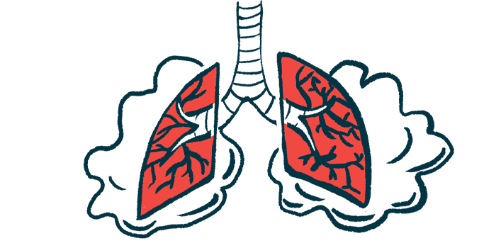
When the mucosa is affected, internal organs, including those involved in breathing, can become damaged. This is well described for the upper respiratory tract — nose, mouth, throat, and voice box — but not so well for the lungs.
In this study, researchers at the Children’s Hospital of Philadelphia reported the cases of three children with generalized JEB who had extensive involvement of the lungs. All three had mutations in the LAMB3 gene.
The first case was a boy who was admitted to the hospital at age 6 months with worsening trouble breathing. He was born with abrasions on the left elbow and multiple fluid-filled blisters and erosions on the face, leading to a diagnosis of JEB.
Microlaryngoscopy and bronchoscopy — two procedures to examine the airways — revealed lesions in the vocal cords and glottis, which is the opening between the vocal cords. A tracheostomy — a surgical opening into the trachea — was performed to provide an airway to the lungs and help with breathing.
He was slowly weaned off breathing support and placed on continuous positive airway pressure (CPAP) therapy to receive constant and steady air pressure. However, his levels of oxygen in the blood dropped, and those of carbon dioxide went up. He also developed recurrent pneumothoraces (collpased lung), which occurs when air leaks into the space between the lungs and the chest wall. Imaging exams revealed widespread lesions in the chest.
At age 7 months, doctors decided to stop supporting the boy’s breathing given the level of disease and the associated complications. Examination of the airways and lungs after death revealed changes to the cells of the bronchi (the tubes connecting the windpipe to the lungs), a buildup of inflammatory cells, and infection (pneumonia).
The second case was a girl aged 1.5 months who presented with a history of noisy breathing, cough, and blood-tinged saliva. She had been diagnosed with JEB in the first weeks of life.
During her hospital stay, she was found to have a low volume of circulating blood. She had ulcers (sores) in the upper part of the voice box, and the vocal cords appeared blunt. She was placed on mechanical ventilation for one week to help with breathing. However, one week later, her breathing problems got worse.
Chest scans revealed a collapse of part of the lungs. Microlaryngoscopy and bronchoscopy revealed inflammation and swelling of the voice box, which narrowed the opening between the vocal cords.
She died at the age of 3 months after an episode of low oxygen levels in the blood. Examination of the airways and lungs after death revealed ulcers, a buildup of inflammatory cells, and enlarged airspaces.
The third case was a girl with a diagnosis of generalized severe JEB who had been admitted to the hospital at 3 months of age for wheezing. At 4.5 months, she was again admitted for trouble breathing with stridor, a harsh, vibrating noise when breathing caused by an obstruction. She also had extensive skin blistering.
Bronchoscopy revealed scarring and softening of the tissues of the voice box and narrowing of its upper part. At 9 months, she presented with trouble breathing and fever. She experienced several episodes of bleeding and septic shock, which occurs when a bodywide infection leads to dangerously low blood pressure. She died at age 1 year.
Examination of the airways revealed scarring of the mucosa lining the voice box and the trachea, which also was inflamed. The lungs had signs of bleeding and pneumonia.
In the three cases, “exacerbation of respiratory distress was associated with mucosal damage,” the researchers wrote.
“These cases illustrate the severe lower airway and parenchymal [functional tissue] complications that can occur in JEB in addition to the more commonly recognized upper airway mucosal involvement,” they wrote. “A multidisciplinary approach that includes families in respiratory management decisions is paramount, as prolonged ventilatory support in generalized severe JEB is generally not well tolerated.”


Leave a comment
Fill in the required fields to post. Your email address will not be published.