DNA, Protein Analyses Improve Diagnosis of Unusual Epidermolysis Bullosa Cases, Study Shows

Combining genetic and protein analyses is a successful strategy to identify unusual cases of epidermolysis bullosa (EB), a German study shows.
The research, “The Position of Targeted Next-generation Sequencing in Epidermolysis Bullosa Diagnosis,” was published in the journal Acta Dermato-Venereologica.
EB is a group of disorders with clinical and genetic variability, collectively characterized by skin fragility and blistering. Although the genetic basis of the disease is well-known, new mutations linked to EB are still being identified.
Mutation analysis is a key element to classifying the disease into its four types and over 30 subtypes. Precise classification is essential for early prognosis (including prenatal), disease management, and genetic counseling.
Unlike in cases of classical EB, molecular and genetic assessment is necessary for a correct diagnosis in newborns, infants, and individuals with moderate or mild skin fragility.
In this study, researchers assessed the molecular pathology of 40 suspected cases of EB.
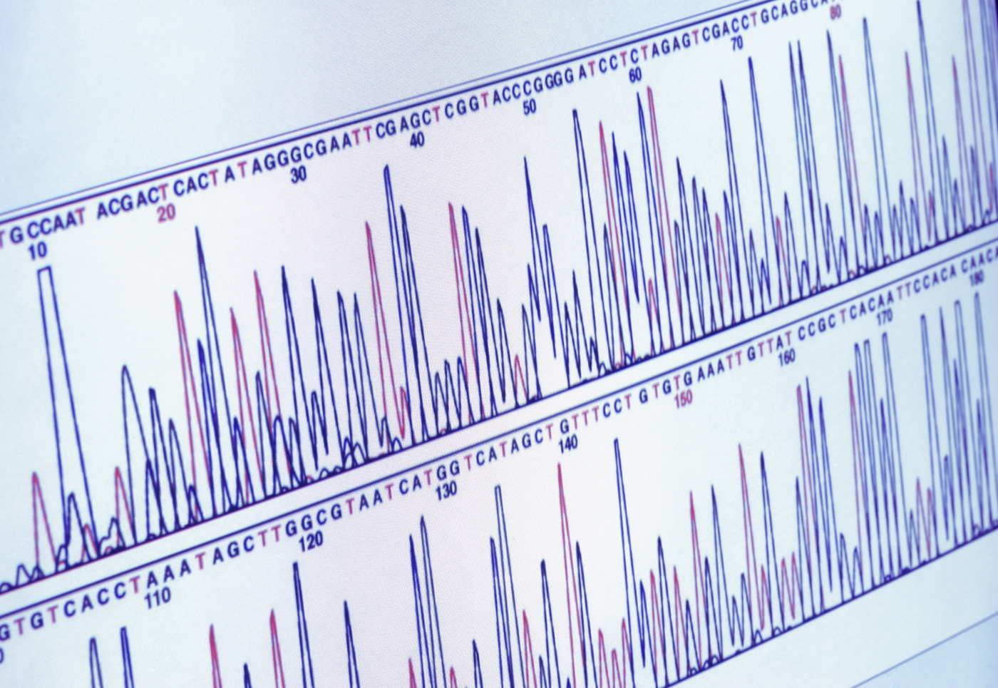
Patients were analyzed by immunofluorescence mapping — IFM, a technique that uses antibodies that bind to proteins in the dermal-epidermal junction zone to assess skin damage — and by targeted next-generation sequencing (NGS), an analysis of multiple DNA sequences. Patients were analyzed beginning in July 2016, when diagnosis with NGS was approved in Germany.
IFM was used to analyze skin biopsy in 25 individuals, and correctly determined EB subtype in 19 cases (76 percent). This technique helped achieve a fast diagnosis in newborns, and helped to determine the consequences of mutations with unknown significance, the researchers observed.
NGS analysis enabled complete elucidation of the molecular pathology in 36 patients (90 percent of the cohort analyzed), including cases with unclear clinical and IFM results. NGS analyzed a panel of 49 genes, 19 of which (if mutated) were known to be associated with EB, while the other 30 genes were related to skin adhesion or to potential genetic modifiers.
Results showed that 12 cases were EB simplex, four were junctional EB, and 15 were dystrophic EB. The most common cause of EB in the study were mutations in the COL7A1 gene, which led to dystrophic EB, one of the major forms of the disease.
Among the identified mutations, 13 had not been previously linked with EB, including two in the KRT5 gene and six in the COL7A1 gene.
The data also showed several mutations not causing EB, but with a potentially modifying role in the disease.
“Based on these preliminary data, we believe that complex constellations of disease-causing mutations and genetic modifiers occur in individual situations,” the investigators wrote. These mutations should be screened in cases of variable EB manifestations in the same family, they advised.
Overall, “this study underscores the efficacy of the strategy of combining targeted NGS with IFM in resolving unusual phenotypes,” the researchers concluded. “It also suggests that, despite technological advances, careful clinical evaluation and deep phenotyping remain crucial factors for successfully establishing EB diagnosis.”


