Cord Blood Platelet Gel Leads to Faster Wound Healing in RDEB Children, Study Says

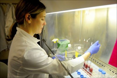
Kate Stringaris, a researcher with the NIH's National Heart, Lung and Blood Institute in Bethesda, Maryland, separates lymphocytes from a blood sample as part of an investigation into natural killer immune cells.
A gel made from platelets — tiny cells involved in blood clotting — that were collected from umbilical cord blood was better and faster at promoting wound healing in children with recessive dystrophic epidermolysis bullosa (RDEB) than a standard approach, a pilot study found.
The children were treated with the gel or with standard therapies after undergoing hand surgery.
The study, “Efficacy of Allogeneic Cord Blood Platelet Gel on Wounds of Dystrophic Epidermolysis Bullosa Patients after Pseudosyndactyly Surgery,” was published in the International Journal of Tissue Repair and Regeneration.
RDEB is caused by mutations in the COL7A1 gene, which contains instructions for making a key protein for skin integrity, called type VII collagen (COL7).
The rare disease is characterized by skin fragility and blistering, with patients developing hand deformities, impaired hand movements, and pseudosyndactyly, a condition in which the fingers or toes appear “fused” by a skin membrane. Pseudosyndactyly can be corrected by surgery, but it is important to prevent the formation of new blisters to avoid further complications, such as infections. Traditionally, this is done with wound dressings, such as gel or hydrogel, while topical antibiotics are used to prevent infections.
Now, researchers at the Skin and Stem Cell Research Center at Tehran University of Medical Sciences, in Iran, investigated the therapeutic effects of an allogeneic platelet gel derived from umbilical cord blood. Notably, allogeneic means derived from a donor.
The study involved 17 children with RDEB, ages 3 to 7, who underwent corrective surgery for pseudosyndactyly in a hand. A total of 14 of the children received the platelet gel, while three were assigned the standard treatment of paraffin gauze wound dressing plus topical antibiotics, and served as controls. Both approaches were given immediately after surgery.
Five days after the first administration of the platelet gel, the wounds were sterilized and washed with a saline solution. Wound surface area and healing progress were then evaluated before applying the gel for a second time. The gel-application process was monitored by a plastic surgeon and two dermatologists, and conducted regularly throughout the study.
Results showed that the wound surface area was reduced by 2 to 4 square centimeters (cm2) after each session in those given the platelet gel, independently of the children’s age or sex. Meanwhile, in the control group, the reduction was 1 to 1.5 cm2.
The healing process, assessed by the tissue’s granulation — the scar tissue formed at the surface of a wound when it is healing — and repair, lasted for 14 to 21 days in children given the platelet gel It took longer, from 35 to 40 days, in the children in the control group.
The researchers also evaluated whether the platelet gel was beneficial for pain management. At each treatment session, they evaluated pain by giving a score according to the patients’ facial symptoms and answers during dressing changing. The findings showed that the level of pain decreased in the platelet gel group compared with those in the control group.
“The current study demonstrated that this gel dressing provided an effective treatment in faster rate of epithelialization [generation of new skin cells] and healing of the wounds, decreased patients’ pain level and post-surgical recovery period, which altogether led to improvements in patients’ overall quality of life,” the scientists concluded.


