
Collagen XVII Mutation Behind Skin Blistering Seen in Mild JEB, Study Confirms
Written by |
The collagen XVII mutation p.R1303Q that causes late-onset, mild junctional epidermolysis bullosa (JEB) is characterized by a weaker collagen XVII connection to another adhesion molecule in the skin, called laminin-332, leading to blistering and thinning skin, a support study says.
The study, “Amino acid substitution in the C-terminal domain of collagen XVII reduces laminin-332 interaction causing mild skin fragility with atrophic scarring,” was published in the journal Matrix Biology.
JEB is caused by defects in genes important to the formation of anchoring filaments and hemidesmosomes — cell fibers and small structures at the skin cell’s surface that work to keep the epidermis (the outermost layer of the skin) attached to the dermis (the underlying layer). These defects can lead to the separation of skin layers and formation of blisters whenever there is friction or trauma.
The researchers studied the cellular and molecular alterations behind JEB in a patient who carried the p.R1303Q mutation in the COL17A1 gene.
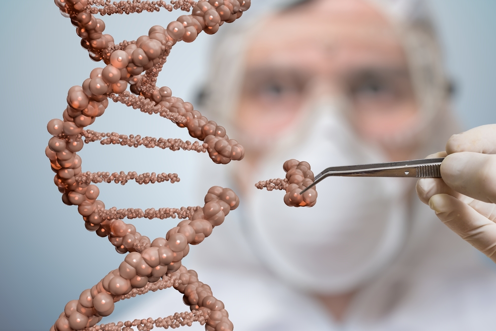
This gene drives the production of protein needed for the formation of collagen type XVII, a major component of hemidesmosomes. This protein crosses the cell membrane of keratinocytes, the predominant cells in the epidermis, extending to outside. The portion of collagen XVII placed outside keratinocytes binds to other adhesion molecules (e.g. laminins and integrins), forming an anchor that holds skin layers together.
“Hemidesmosomal collagen XVII essentially contributes to cell adhesion” and regulates skin cell “directionality and proliferation during skin regeneration,” researchers said.
The mutation under study, called p.R1303Q, leads to the formation of a modified collagen XVII associated with a mild, late-onset JEB.
Prior research showed that this mutation did not change collagen XVII levels, but patients having it “developed late-onset skin blistering, progressive skin atrophy, loss of dermatoglyphs [fingerprints, lines, mounts and shapes of hands], scarring and nail anomalies,” researchers said.
Yet little is known about the mechanisms involved in these skin manifestations underlain by such a mutation. To find answers, researchers cultured in a laboratory keratinocytes from a patient with JEB carrying the p.R1303Q mutation.
After performing several experiments with these cells, scientists demonstrated that this particular mutation affected the end portion of collagen XVII, the one located outside keratinocytes and bridging these cells’ attachment to the dermis.
The mutation changed the shape and chemical properties of collagen XVII, decreasing its ability to bind laminin-332, a matrix protein deposited outside cells and another important component for skin attachment.
Researchers observed that although the mutant collagen XVII was expressed at normal levels and was correctly placed at the cell membrane of keratinocytes, its defective binding to laminin-332 seemed to turn keratinocytes ”less adhesive,” detaching more easily. They also “showed migratory defects” and higher rates of cell death, compared to cells with normal collagen.
The findings suggest that such defects in collagen molecular interactions in turn altered keratinocytes’ behavior and “led to blister formation, delayed wound healing and skin [thinning].”
“Thus, collagen XVII-laminin interactions are important for keratinocyte adhesion, motility and survival, which in turn promote skin integrity, wound healing and protect against skin aging,” researchers said.


Leave a comment
Fill in the required fields to post. Your email address will not be published.